Full Financial Member
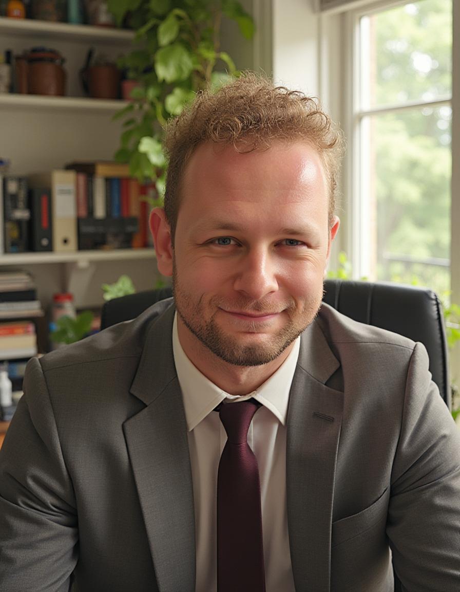
Thomas A. Stratton
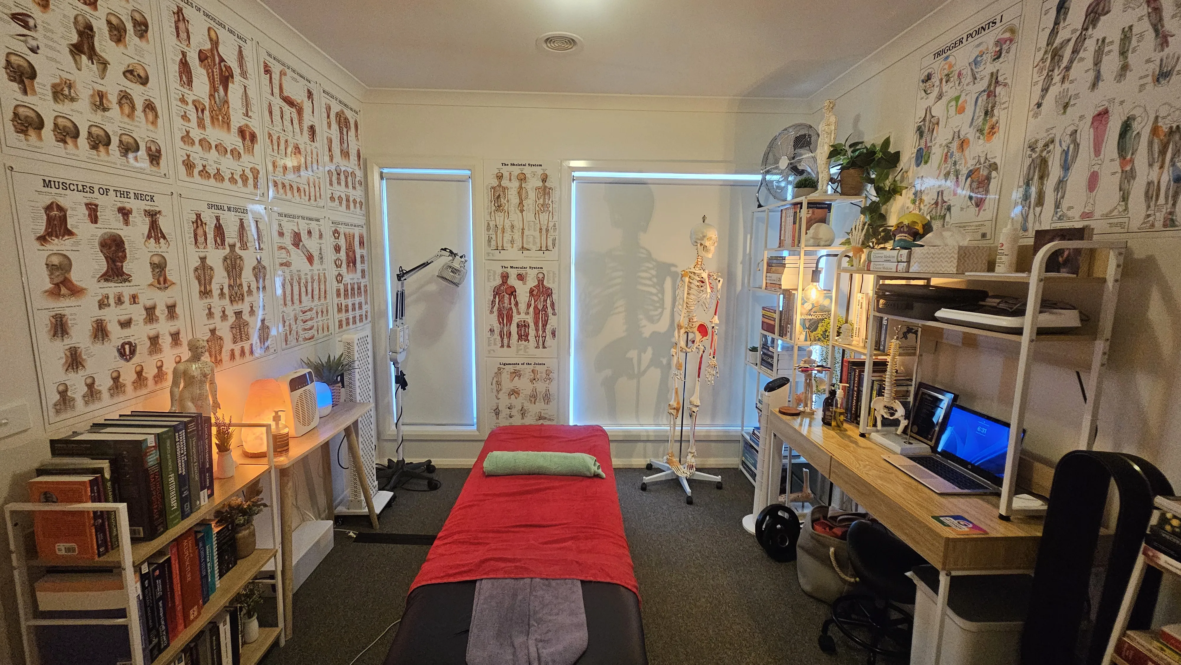
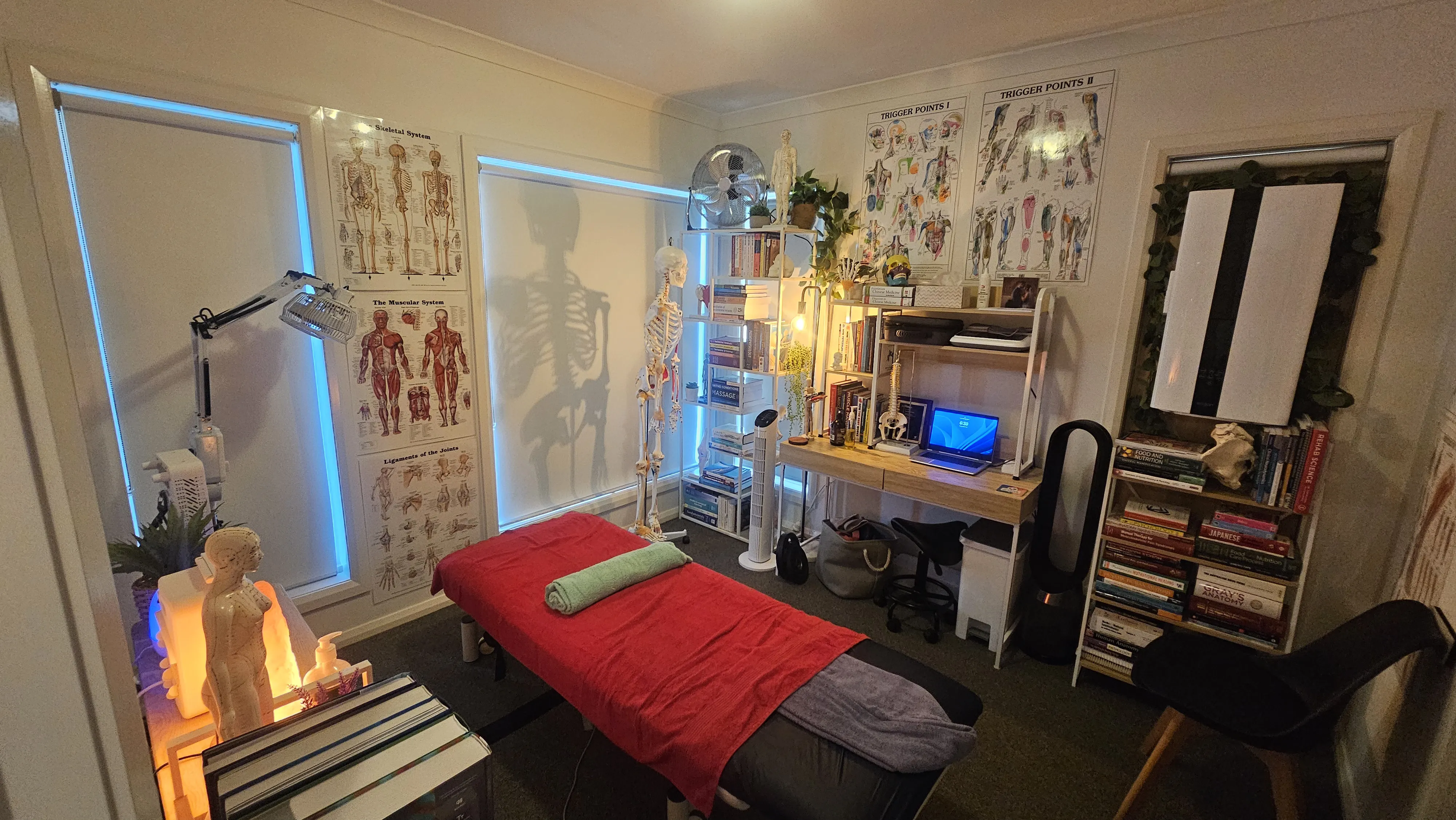
A qualified Remedial Massage Therapist and Myotherapist committed to evidence-based clinical practice. My treatment philosophy is informed by current research in pain neuroscience, musculoskeletal pathology, and rehabilitation science.
My clinical practice draws on a broad scope of training across multiple institutions, encompassing over 600 hours of continuing professional development in dry needling alone. This includes advanced certifications in Huneke neural therapy, electro-dry needling, fascial and scar tissue techniques, pregnancy massage, Gua Sha, electrotherapy, IASTM, and manual therapy approaches including Maitland and Mulligan mobilisation techniques.
Whether you are managing acute musculoskeletal injury, chronic pain, neuromuscular dysfunction, postural imbalance, or seeking proactive maintenance care, I apply the same rigorous, evidence-informed approach to every session.
Qualifications & Certifications
SAGE Institute & The Gordon
Melbourne Institute of Massage & Myotherapy
Pregnancy Massage Australia
Advanced Huneke, Scar Tissue & Fascial Techniques
Health Traditions
RockTape Australia
RockTape Australia
Advanced Clinical Education
Pain Science Informed
Treatment integrates current understanding of central sensitisation, nociception, and neuroplasticity to address both peripheral and central drivers of pain.
600+ Hours Dry Needling
Extensive post-graduate training including Huneke neural therapy, fascial needling, scar tissue techniques, and electro-dry needling across multiple accredited providers.
Outcome-Focused
Systematic reassessment protocols track progress and adapt treatment plans to ensure measurable, clinically meaningful improvement at every stage.